Laparoscopic Appendicectomy
General Surgery · MRCS Part B · Core Trainee
Operative Simulator
Work through the procedure step by step. Tick every item on each checklist to unlock the next step.
Operative Guide
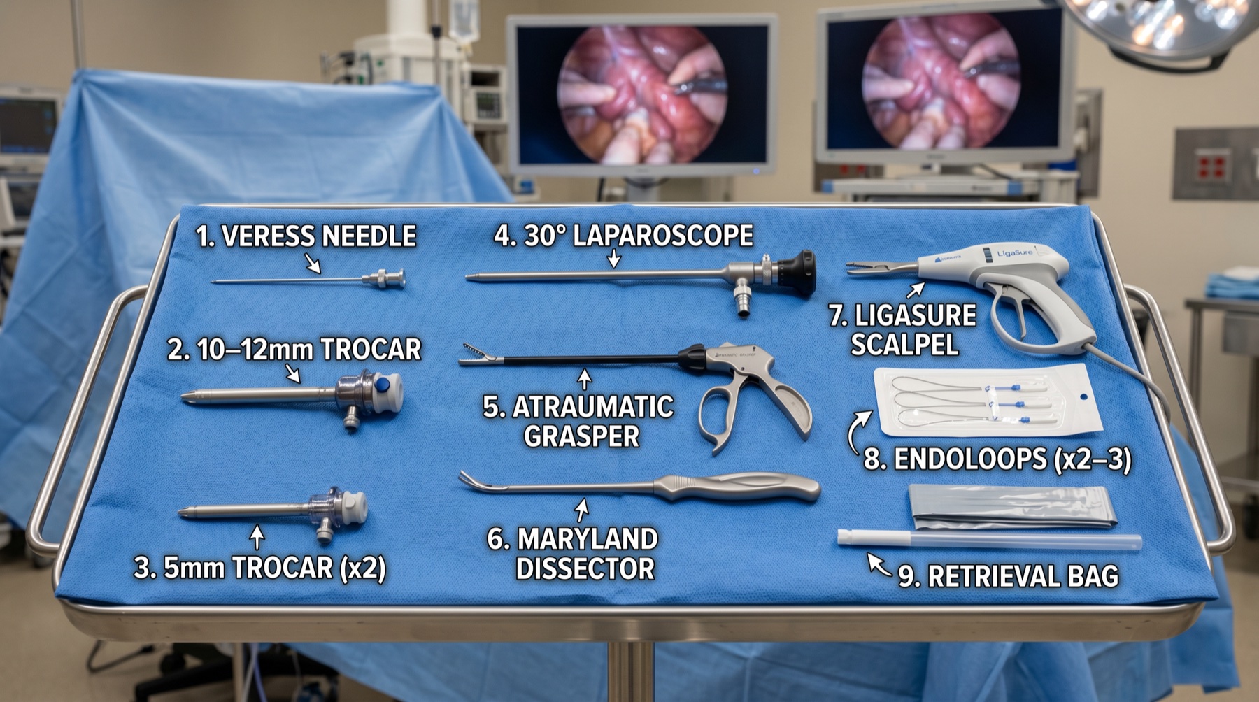
Instruments
Anatomy Danger Zones
Appendicular artery
A branch of the ileocolic artery. If not secured before division it retracts into the mesoappendix and causes significant bleeding.
Inferior epigastric vessels
Run in the anterior abdominal wall. Insert working ports lateral to these vessels, transilluminating where possible.
Right ureter
Crosses over the pelvic brim. At risk during deep or retrocaecal dissection, identify and protect it.
Iliac vessels
Lie deep to the operative field. At risk with Veress entry and with deep dissection in the pelvis.
Complications
Early
- •Wound / port-site infection (~5%)
- •Intra-abdominal collection (3–5%)
- •Ileus, common, usually self-limiting
- •Bleeding, rare
- •Bowel injury, rare
- •Stump leak, rare
Late
- •Port-site hernia (if fascia not closed)
- •Adhesional bowel obstruction
- •Chronic right iliac fossa pain
- •Stump appendicitis
- •Carcinoid (0.3%), if >2cm refer to colorectal MDT for right hemicolectomy
MCQ Quiz
Where must the endoloops be placed when securing the appendix base?
You find a macroscopically normal appendix in a woman undergoing laparoscopy for suspected appendicitis. What do you do?
The appendicular artery is divided before haemostasis is achieved and it retracts. What is the main risk?
Histology reports a 2.4 cm carcinoid (neuroendocrine tumour) of the appendix. What is the next step?
The appendix base is necrotic and friable. What is the best method of control?
Post-op Care
Immediate
- ›Paracetamol + ibuprofen for analgesia
- ›Early mobilisation
- ›IV antibiotics 24h if perforated, then switch to oral
- ›Monitor for pyrexia after day 3 (suggests collection)
Discharge
- ›Uncomplicated: same day or next day
- ›Perforated: 2–4 days
- ›Oral co-amoxiclav 5 days if perforated
- ›Safety-net advice on collection / wound infection
Follow-up
- ›Histology review for all specimens
- ›Carcinoid / neoplasm referral if indicated
- ›GP wound check
Sick note
- ›Office work: 1–2 weeks
- ›Manual work: 4–6 weeks
- ›No heavy lifting for 4–6 weeks