Endoscopic Classifications — A Working Reference
For the surgical registrar in the endoscopy suite and the clinic.
This is a reference for the classifications you will be asked about on the list, and — more importantly — what each one actually changes about management. A classification you can recite but not act on is worth very little.
Part One opens with how to perform a complete diagnostic OGD, and also covers hiatus hernia, peptic ulcer disease and H. pylori: the endoscopy knowledge a general surgeon is expected to have.
Last reviewed: October 2026. Guidelines change. Always check your trust protocol and the current BSG/ESGE/NICE position before acting on anything here.
PART ONE — UPPER GI ENDOSCOPY (OGD)
1. Performing a Diagnostic OGD: Technique and Systematic Survey
Classifications are only useful if you have seen everything. This is a practical walk-through of a complete diagnostic OGD, from intubation to withdrawal.
Set-up
- Position: left lateral, head on a small firm pillow in a neutral position, neck slightly flexed. IV access ideally in the right arm. Monitoring, supplemental oxygen and a biteguard in place before you start.
- Scope checks before intubation: tip angulation, air and water, suction and image quality (white balance if needed). Lubricate the distal tip.
- Grip: the control head sits in the left palm. Left thumb on the up/down wheel, index finger on the air/water and suction buttons, middle finger helping the thumb on big movements. The right hand pushes, pulls and applies torque.
- Keep the shaft straight from the mouth to your hands. Loops in the shaft absorb torque, so rotation stops reaching the tip and you lose orientation.
Intubation under direct vision
- Hold the shaft at about 30 cm and rehearse the up/down movement so the tip will follow the curve of the pharynx.
- Pass over the tongue watching the patient, then switch to the monitor. The image is inverted at this stage.
- Stay in the midline by following the median groove of the tongue. The uvula appears briefly, then the epiglottis.
- The cricoarytenoid cartilage and vocal cords sit at the top of the screen. Angle down behind the cartilage and pass slightly to one side of the midline, because the cartilage bulges centrally against the cervical spine.
- A red-out means the tip is against the closed cricopharyngeus. Insufflate, keep gentle steady pressure and ask the patient to swallow. It should slip through within a few seconds.
- Watch the mucosa slide past in the upper oesophagus. This is where a Zenker's diverticulum can swallow the scope.
If the view is lost, the teeth come into view or the tongue deflects the tip, withdraw and start again. Force is never needed.
Two golden rules: do not push if you cannot see; if in doubt, inflate and pull back.
Oesophageal landmarks (adult, distance from incisors)
| Distance | Landmark |
|---|---|
| ~15 cm | Cricopharyngeus |
| ~25 cm | Aortic arch and left main bronchus (extrinsic indentation) |
| ~35 cm | Left atrium (visible pulsation) |
| ~38-40 cm | GOJ: top of the gastric folds |
Ask the patient to sniff to show the diaphragmatic pinch, and record its distance separately from the GOJ and the Z-line. These three numbers are what let you diagnose a hiatus hernia or Barrett's (see the hiatus hernia section below).
Entering the stomach
- Through the cardia the tip meets the posterior wall of the lesser curve. Rotate anticlockwise (left), deflect slightly down and add air.
- Turning right here curls the tip back into the fundus. This accidental retroflexion is the commonest way to get lost. If in doubt: withdraw, inflate, turn sharply left.
- Orientation in the left lateral position, buttons up: lesser curve on the right, longitudinal greater curve folds on the left (usually with a fluid pool), posterior wall below, angulus in the distance.
- Aspirate the resting juice to reduce aspiration risk. Flush simethicone down the channel if there is a lot of foam.
- To reach the antrum: tip up and rotate clockwise (about 90°). The distended stomach is a J-shape draped over the spine, and the scope follows it in a clockwise spiral. Then deflect slightly down to line up with the pylorus.
- Watch antral and pyloric peristalsis. Asymmetry of a contraction wave suggests current or previous disease, and poor motility is a clue to linitis plastica.
Pylorus and duodenum
- Approach with the pylorus centred and push gently. Deflating a little or angling down can help a tight or deformed pylorus. Give IV hyoscine butylbromide or glucagon if motility is a problem.
- Once through, the gastric loop straightens and the tip jumps to the distal bulb. Pull back a few centimetres and insufflate to see the whole bulb. The inferior bulb just beyond the pyloric ring is the area most often missed.
- Into D2: at the superior duodenal angle, rotate right about 90°, tip right, tip up, then pull back. Pulling straightens the loop along the greater curve and drives the tip forward round the angle. Pushing only enlarges the loop. The papilla is at about 60 cm.
Retroflexion (J manoeuvre)
- Do it after the duodenum so you are not over-inflating on the way in.
- Tip in the mid stomach at or just beyond the angulus, insufflate, then tip up 180° using both wheels. Withdraw and rotate to see the lesser curve, cardia and fundus. This is where you assess the Hill grade and any hiatus hernia from below.
- Do not pull back far enough to impact the retroflexed tip in the oesophagus. Return the wheels to neutral afterwards.
- A lax cardia that will not hold air: roll the patient slightly towards supine.
Withdrawal
- Re-inspect everything on the way out. The proximal lesser curve is a recognised blind spot.
- Aspirate all air and fluid from the stomach before removing the scope.
- BSG/AUGIS quality standards: photo-document the key landmarks, and aim for an inspection time of at least 7 minutes for a first diagnostic OGD (and for gastric IM surveillance).
When things go wrong
- Patient distress: stop if the cause is not obvious and fixable. Severe pain is rare and suggests a complication (perforation or a cardiac event). Tachycardia and bradycardia can both signal distress.
- Food in the stomach despite fasting = gastric outlet obstruction until proven otherwise. Scope channels block quickly and the aspiration risk is real. You can slide along the lesser curve over the food to look for a distal lesion, but usually it is wiser to stop, lavage and repeat.
- Poor view: the lens is against mucosa or covered. Withdraw, insufflate, wash. Check the air pump, connections and O-rings, because inadequate insufflation is the commonest cause of getting lost in normal anatomy.
- Post-surgical stomach: hyperaemia and bile are normal after partial gastrectomy or gastroenterostomy. There is a higher cancer risk in the remnant, especially near the stoma, so biopsy generously within 3 cm of it if suspicious. Marginal ulcers sit at or just beyond the anastomosis.
Biopsy technique
- Approach the lesion face-on, keep the forceps close to the tip and move the scope to the target rather than pushing the forceps out.
- In the oesophagus, angle the tip against the wall with the forceps barely protruding. Spiked forceps help with tangential targets.
- Ulcers: biopsy the rim in all four quadrants. The base gives slough, unless you suspect a viral cause (CMV, HSV).
- Tumours: avoid necrotic areas, which give non-diagnostic samples.
- Submucosal lesions: standard biopsies are usually normal because the forceps do not cross the muscularis mucosae (neuroendocrine tumours are the exception). Use EUS-FNA or a snare biopsy.
- Brush cytology is useful in tight strictures where biopsies are hard to get, and for candida.
- If a biopsy site bleeds and obscures the lesion, wash with water or dilute adrenaline.
- Label pots by site, state the clinical question, and say on the form if you want H. pylori looked for. Some labs will not stain for it otherwise.
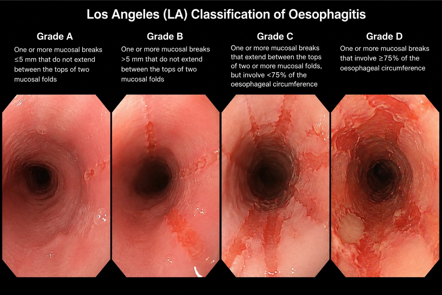
2. Los Angeles (LA) Classification — Reflux Oesophagitis
The classification you will be asked for most often. It grades mucosal breaks, not erythema.
| Grade | Definition |
|---|---|
| A | One or more mucosal breaks ≤5 mm, not extending between the tops of two mucosal folds |
| B | At least one mucosal break >5 mm, not extending between the tops of two mucosal folds |
| C | Mucosal break continuous between the tops of two or more mucosal folds, involving <75% of the circumference |
| D | Mucosal break involving ≥75% of the oesophageal circumference |
What it changes
- A is of doubtful clinical significance — found in a meaningful proportion of asymptomatic people. Do not over-interpret it.
- B confirms reflux disease. PPI therapy.
- C and D are severe oesophagitis. Two consequences:
- Barrett's cannot be reliably excluded in the presence of severe inflammation. Repeat OGD after 8 weeks of high-dose PPI to reassess for Barrett's and to exclude an underlying malignancy.
- Long-term maintenance PPI is indicated; these patients rarely stay healed off treatment.
- Severe oesophagitis also raises the question of eosinophilic oesophagitis, pill oesophagitis, infective oesophagitis (candida, CMV, HSV) and caustic injury — the pattern and history distinguish them.
Practical points
- Requires mucosal break (erosion or ulcer with a clear edge). Erythema, oedema and increased vascularity alone do not qualify.
- Supersedes the older Savary-Miller classification (grades I–IV), which is still occasionally quoted by older consultants — I–II roughly maps to LA A–B, III to LA C–D, IV adds complications (stricture, ulcer, Barrett's).
- A normal OGD does not exclude GORD — most reflux disease is non-erosive (NERD).
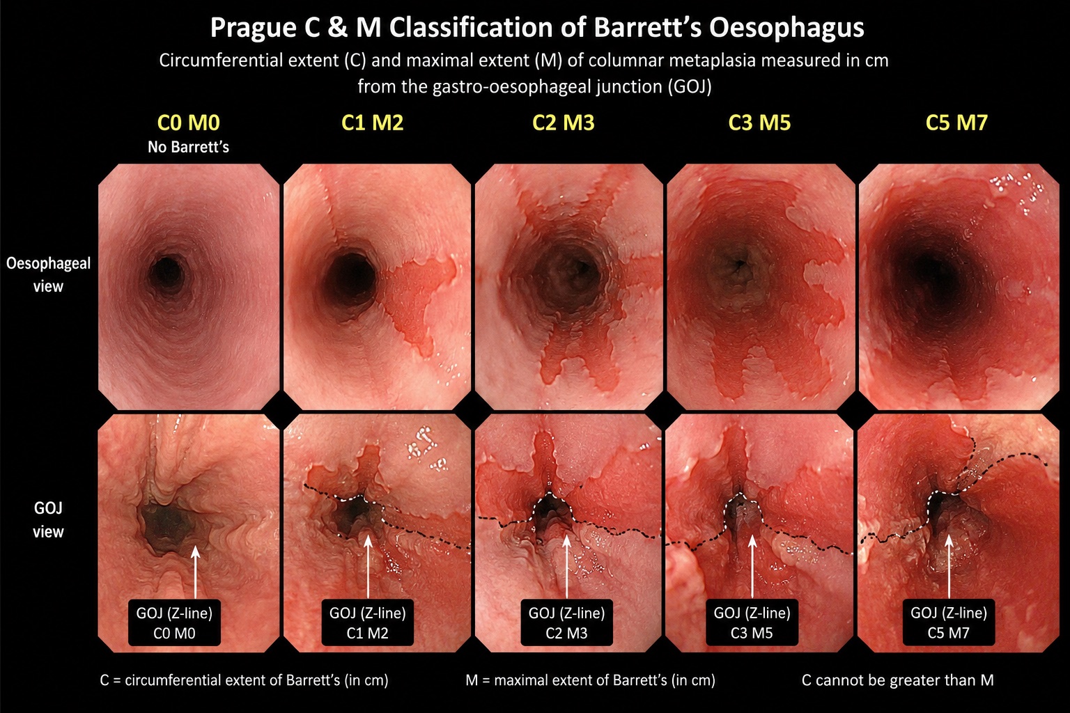
3. Prague C&M Criteria — Barrett's Oesophagus
The standard for reporting the extent of a Barrett's segment.
- C = Circumferential extent in cm above the GOJ
- M = Maximal extent in cm above the GOJ
Both measured from the top of the gastric folds (which defines the GOJ endoscopically), to the proximal limit of columnar-lined mucosa.
Example: C2M5 = 2 cm of circumferential columnar mucosa, with a tongue extending to 5 cm.
Rules
- M is always ≥ C.
- Islands of columnar mucosa proximal to the main segment are described separately and are not included in M.
- Report the position of the diaphragmatic hiatus separately — this identifies a hiatus hernia and prevents mistaking hernia for Barrett's.
- A segment must be ≥1 cm to be called Barrett's. Below this it is an irregular Z-line, which should not be biopsied routinely and does not enter surveillance.
UK vs US definition — worth knowing
- BSG (UK): diagnosis requires ≥1 cm of columnar-lined oesophagus with histological confirmation of columnar metaplasia. Intestinal metaplasia is not mandatory, though its presence increases risk.
- ACG/AGA (US): intestinal metaplasia with goblet cells is required.
This is a classic viva discriminator.
Biopsy — the Seattle protocol
- Targeted biopsies of any visible lesion first (separate pots), then
- Four-quadrant random biopsies every 2 cm of segment length
- Every 1 cm if dysplasia is known or suspected
Adherence to the Seattle protocol in real-world practice is poor — audits repeatedly show around half of surveillance endoscopies comply. This is a very easy audit project if you need one.
Surveillance intervals
BSG (UK practice):
| Finding | Interval |
|---|---|
| Segment <3 cm, no intestinal metaplasia | Repeat to confirm; consider discharge |
| Segment <3 cm with intestinal metaplasia | 3–5 years |
| Segment ≥3 cm | 2–3 years |
ESGE 2023 (increasingly influential, some UK units have adopted it):
| Segment length | Interval |
|---|---|
| ≥1 cm and <3 cm | 5 years |
| ≥3 cm and <10 cm | 3 years |
| ≥10 cm | Refer to a Barrett's expert centre |
ESGE also suggests considering stopping surveillance at age 75 if no dysplasia has developed.
Dysplasia
- Indefinite for dysplasia → optimise PPI, repeat at 6 months.
- Low-grade dysplasia → confirm with a second expert GI pathologist (inter-observer agreement is poor and a large proportion are downgraded). If confirmed, endoscopic ablation (RFA) is now preferred over surveillance.
- High-grade dysplasia or intramucosal cancer → endoscopic resection of any visible lesion for accurate staging, then ablation of residual Barrett's. Refer to a specialist centre.
4. Paris Classification — Superficial Neoplastic Lesions
Applies throughout the GI tract — oesophagus, stomach, colon. Describes morphology, which predicts depth of invasion.
Type 0 = superficial lesion (endoscopic appearance suggests invasion no deeper than submucosa)
| Subtype | Description |
|---|---|
| 0-Ip | Protruding, pedunculated |
| 0-Is | Protruding, sessile |
| 0-IIa | Slightly elevated (<2.5 mm) |
| 0-IIb | Completely flat |
| 0-IIc | Slightly depressed |
| 0-III | Excavated / ulcerated |
Mixed types are common and are written with the dominant component first: 0-IIa+IIc, 0-Is+IIc.
The 2.5 mm rule
The elevated/flat cut-off is the height of closed biopsy forceps (approximately 2.5 mm). Hold the forceps beside the lesion — this is the practical bedside method and worth knowing when asked how you decided.
Why it matters
Depressed and excavated components (IIc, III) carry a substantially higher risk of deep submucosal invasion — these are the lesions you must not attempt to snare blindly. A 0-IIa+IIc lesion is far more concerning than a 0-IIa of the same size.
Combine with the non-lifting sign (failure of the lesion to lift on submucosal injection), which suggests deep invasion or fibrosis and contraindicates endoscopic mucosal resection.
5. Forrest Classification — Bleeding Peptic Ulcer
The single most clinically actionable endoscopic classification. Know it cold.
| Class | Appearance | Rebleed risk (untreated) | Endoscopic therapy? |
|---|---|---|---|
| Ia | Spurting arterial bleeding | ~55–90% | Yes |
| Ib | Oozing bleeding | ~50% | Yes |
| IIa | Non-bleeding visible vessel | ~43% | Yes |
| IIb | Adherent clot | ~22% | Consider — see below |
| IIc | Flat pigmented spot | ~10% | No |
| III | Clean ulcer base | ~5% | No |
What it changes
- Ia, Ib, IIa = high risk. Endoscopic haemostasis is mandatory. Adrenaline injection alone is insufficient — always combine with a second modality: mechanical (through-the-scope clips, over-the-scope clips), thermal (bipolar coagulation, heater probe), or topical haemostatic powder. Follow with high-dose IV PPI infusion (80 mg bolus then 8 mg/hr for 72 hours).
- IIb (adherent clot) is the contentious one. Reasonable practice is vigorous irrigation or cold-snare removal of the clot to reveal the underlying stigmata, then treat accordingly. If the clot will not shift, treat as high risk.
- IIc and III = low risk. No endoscopic therapy. Oral PPI, early feeding, and these patients can often be discharged early — a Rockall score helps.
Adjuncts worth mentioning
- Pre-endoscopy IV erythromycin (250 mg, 30–120 min before) as a prokinetic improves gastric visualisation and reduces the need for repeat endoscopy.
- Rebleeding after successful therapy → repeat endoscopy first. Second failure → interventional radiology embolisation in preference to surgery in most units.
- Surgery (under-running for duodenal ulcer via duodenotomy; partial gastrectomy rarely) for failure of endoscopic and radiological control.
- Always biopsy gastric ulcers and arrange repeat OGD at 6–8 weeks to confirm healing and exclude malignancy. Duodenal ulcers do not require this.
- Test and treat H. pylori in all peptic ulcer disease.
6. Glasgow-Blatchford and Rockall Scores
Not endoscopic classifications as such, but you will be asked alongside Forrest.
Glasgow-Blatchford (pre-endoscopy) — urea, haemoglobin, systolic BP, pulse, melaena, syncope, hepatic disease, cardiac failure.
- Score 0–1 → very low risk, consider outpatient management with early OGD.
- Score ≥6 → high risk, needs inpatient endoscopy within 24 hours.
Rockall (full score, post-endoscopy) — age, shock, comorbidity, diagnosis, stigmata of recent haemorrhage. Predicts rebleeding and mortality.
7. Peptic Ulcer Disease: Endoscopic and Surgical Management
Forrest tells you what to do with a bleeding ulcer today. This section covers everything else: what caused it, whether it might be cancer, and when it becomes a surgical problem.
Causes
- H. pylori (historically around 90% of DUs and 70% of GUs; the proportion is falling)
- NSAIDs and aspirin, including over-the-counter use the patient does not mention
- Less common: malignancy, Zollinger-Ellison syndrome, Cameron lesions in a large hiatus hernia, Crohn's disease, CMV, cocaine, and stress ulceration in critically ill patients
Johnson classification of gastric ulcers (a surgical viva favourite)
| Type | Site | Acid |
|---|---|---|
| I | Lesser curve at the incisura (most common) | Low |
| II | Body ulcer plus a duodenal ulcer | High |
| III | Prepyloric (behaves like a DU) | High |
| IV | High on the lesser curve near the GOJ | Low |
| V | Anywhere, NSAID-related | Either |
Gastric vs duodenal ulcer at OGD
| Gastric ulcer | Duodenal ulcer | |
|---|---|---|
| Malignancy risk | Yes, so every GU is biopsied | Very low, so the ulcer itself is not routinely biopsied |
| Biopsy | Multiple samples from the rim in all quadrants, plus H. pylori biopsies | H. pylori biopsies (antrum, plus body if on PPI) |
| Treatment | Full-dose PPI for 8 weeks; stop NSAIDs; eradicate H. pylori | Full-dose PPI for 4-8 weeks; eradicate H. pylori |
| Follow-up | Repeat OGD at 6-8 weeks (NICE) to confirm healing, re-biopsy and retest H. pylori | No routine repeat OGD |
Benign or malignant?
| Favours benign | Favours malignant |
|---|---|
| Round, punched out, smooth flat edge | Raised, rolled or irregular edge, varying in height |
| Clean base | Nodular, lumpy or haemorrhagic base |
| Folds radiating smoothly to the margin | Folds that club, fuse or stop short of the edge |
| Single, lesser curve at or above the angulus | Surrounding abnormal mucosa |
You cannot reliably tell them apart by appearance, so biopsy every gastric ulcer. Multiple, punched-out, sometimes very large and oddly shaped ulcers suggest NSAIDs. Early gastric cancer can mimic a small benign ulcer, a chronic erosion or a flat polyp.
Duodenal ulcer appearances
- Usually on the anterior or posterior wall of the bulb, often multiple.
- Healing and scarring cause bulbar deformity: a shelf-like fold that can create a pseudodiverticulum.
- Ulcers or duodenitis in D2 suggest Zollinger-Ellison syndrome or pancreatic disease. Small aphthous ulcers in D2 raise Crohn's.
The refractory ulcer: work through this list
- Compliance with PPI
- Persistent H. pylori (false negatives on PPI are common, so retest properly)
- Hidden NSAID or aspirin use
- Smoking
- Malignancy: re-biopsy
- Zollinger-Ellison syndrome: multiple, distal (D2 or beyond) or recurrent ulcers, often with diarrhoea. Check fasting gastrin off PPI.
When it becomes surgical
Bleeding (see Forrest above):
- No PPI before endoscopy for suspected non-variceal bleeding (NICE CG141). Start it after endoscopic haemostasis.
- Endoscopy immediately after resuscitation if unstable, otherwise within 24 hours.
- Rebleed: repeat endoscopy. If that fails, IR embolisation (preferred if unstable) or surgery.
- Posterior DU over the gastroduodenal artery (GDA): this is the classic surgical bleeder. Through a duodenotomy (extended through the pylorus if needed), underrun the GDA with a three-point ligation: sutures above and below the bleeding point, plus a U-stitch medially for the transverse pancreatic branch. Close the pylorus as a pyloroplasty if it was opened.
Perforation:
- Omental (Graham) patch, open or laparoscopic, with washout.
- Conservative management (Taylor approach) for a sealed perforation in a selected patient.
- Always biopsy a perforated gastric ulcer (or re-scope later), and test for and eradicate H. pylori afterwards.
Gastric outlet obstruction:
- Food residue at OGD after an adequate fast. Exclude malignancy first.
- Benign pyloroduodenal stenosis from ulcer scarring can be balloon dilated, but recurrence is common.
- Malignant gastric outlet obstruction responds well to enteral stenting.
8. Helicobacter pylori: Testing and Eradication
The organism
Gram-negative, spiral, flagellated, urease-positive and microaerophilic. It lives in the mucus layer, mainly in the antrum. It is a WHO class I carcinogen.
Associations: duodenal and gastric ulcer, gastric adenocarcinoma (via the Correa cascade), gastric MALT lymphoma (low-grade disease can be cured by eradication alone), iron-deficiency anaemia and ITP.
Testing
| Test | Type | Notes |
|---|---|---|
| Rapid urease test (CLO) | Invasive, at OGD | Biopsy placed in a urea gel with a pH indicator. Urease makes ammonia and the gel turns from yellow to pink/red. Fast and cheap |
| Histology | Invasive | Giemsa or immunohistochemistry. Also grades gastritis, atrophy and IM |
| Culture | Invasive | For antibiotic sensitivities after repeated treatment failure |
| Urea breath test (¹³C) | Non-invasive | Shows active infection. Good test of cure |
| Stool antigen | Non-invasive | Shows active infection. Good test of cure |
| Serology | Non-invasive | Cannot distinguish past from current infection, so never a test of cure |
Washout before testing: stop PPIs for 2 weeks and antibiotics or bismuth for 4 weeks. Otherwise false negatives are common.
At OGD:
- Biopsy the antrum, and add the body or fundus if the patient is on a PPI (the organism migrates proximally).
- During an acute bleed, false negatives are more frequent, so if the result is negative, retest later.
Eradication (NICE first line: 7 days, twice daily)
| Situation | Regimen |
|---|---|
| First line | PPI + amoxicillin 1 g + clarithromycin 500 mg or metronidazole 400 mg |
| Penicillin allergy | PPI + clarithromycin 250 mg + metronidazole 400 mg |
| Second line | PPI + amoxicillin + whichever of clarithromycin/metronidazole was not used first time |
| Further failures | Quinolone- or tetracycline/bismuth-based regimens; consider culture and sensitivity |
International guidance (Maastricht VI) favours 14 days and bismuth quadruple therapy first line where clarithromycin resistance is high. UK practice varies, so check your local antimicrobial guideline.
Test of cure
- Not routine for uncomplicated dyspepsia.
- Do test after a gastric ulcer, a bleeding or complicated ulcer, MALT lymphoma, or if symptoms persist.
- Use a breath or stool test at least 4 weeks after treatment, off PPI for 2 weeks.
9. Gastritis — The Systems That Actually Matter
Your consultant asking about "gastritis appearances" is usually asking one of three things: is this H. pylori?, is this atrophic?, and does this patient need surveillance?
9a. Updated Sydney System — the histological framework
Describes gastritis by topography, morphology and aetiology. Five features graded 0–3 (none / mild / moderate / marked):
- Chronic inflammation
- Neutrophil activity
- Atrophy
- Intestinal metaplasia
- H. pylori density
The Sydney biopsy protocol — 5 biopsies:
- 2 from the antrum (lesser and greater curve, 2–3 cm from pylorus)
- 1 from the incisura angularis
- 2 from the corpus (lesser and greater curve)
Antrum and corpus should go in separate pots — this is what allows OLGA/OLGIM staging. If you send them all in one pot, the pathologist cannot stage.
9b. OLGA and OLGIM — the staging systems
Combine antral and corpus scores into a stage 0–IV.
- OLGA stages atrophy
- OLGIM stages intestinal metaplasia — preferred in practice because inter-observer agreement for intestinal metaplasia is considerably better than for atrophy
Stage III–IV = high risk of gastric cancer and warrants surveillance.
9c. Kimura-Takemoto — the endoscopic atrophic border
Describes how far the atrophic border has advanced from antrum towards fundus.
| Type | Description |
|---|---|
| C-1, C-2, C-3 | Closed type — atrophic border confined to the lesser curve |
| O-1, O-2, O-3 | Open type — border has passed onto the anterior/posterior walls and fundus |
C-3 and all O types = extensive atrophy = higher cancer risk.
9d. Kyoto Classification of Gastritis
Endoscopic scoring system (total 0–8) using five features:
- Atrophy (0–2)
- Intestinal metaplasia (0–2)
- Enlarged folds (0–1)
- Nodularity (0–1)
- Diffuse redness (0–2)
A score of ≥4 suggests significantly elevated gastric cancer risk.
Kyoto also helps classify H. pylori status endoscopically:
- Never infected — regular arrangement of collecting venules (RAC) present, fundic gland polyps, no atrophy
- Current infection — diffuse redness, mucosal swelling, enlarged folds, sticky mucus, nodularity
- Past infection (eradicated) — patchy redness, map-like redness, atrophy without diffuse redness
The presence of RAC in the lower body is a useful bedside sign of an H. pylori-naive stomach.
9e. EGGIM — Endoscopic Grading of Gastric Intestinal Metaplasia
Uses virtual chromoendoscopy (NBI/BLI) to score intestinal metaplasia in 5 areas (2 antrum, 2 corpus, incisura), each 0–2. Total 0–10.
EGGIM ≥5 identifies extensive intestinal metaplasia and correlates with OLGIM III/IV — allowing risk stratification at the time of endoscopy without waiting for histology.
9f. Surveillance — MAPS III (ESGE/EHMSG/ESP)
| Finding | Surveillance |
|---|---|
| Mild–moderate atrophy or IM confined to antrum, no risk factors | No surveillance |
| IM at a single location plus a risk factor (family history of gastric cancer, incomplete-type IM, persistent H. pylori) | 3 years |
| Extensive endoscopic changes (Kimura C-3+ / EGGIM ≥5) or advanced histology (OLGA/OLGIM III–IV) | 3 years |
| The above plus family history of gastric cancer | 1–2 years |
| Gastric dysplasia | Expert centre; low-grade 1 year, high-grade → endoscopic resection |
Eradicate H. pylori in everyone with premalignant gastric conditions — it is the single most effective intervention.
9g. Gastritis Types A, B and C
Gastritis is a histological diagnosis. Endoscopic redness correlates poorly with histology, so describe what you see ("erythematous gastropathy") and biopsy rather than labelling it gastritis.
| Type | Cause | Distribution | Key associations |
|---|---|---|---|
| A: autoimmune | Anti-parietal cell and anti-intrinsic factor antibodies | Body-predominant, antrum spared | B12 deficiency, pernicious anaemia, achlorhydria, raised gastrin, type 1 gastric NETs |
| B: bacterial | H. pylori | Starts in the antrum, progresses to pangastritis | Peptic ulcer, atrophy, IM, adenocarcinoma, MALT lymphoma |
| C: chemical/reactive | NSAIDs, alcohol, bile reflux (after gastrectomy or pyloroplasty) | Antrum or anastomosis | Erosions, foveolar hyperplasia |
Others: lymphocytic (associated with coeliac disease), eosinophilic, granulomatous (Crohn's, sarcoid) and CMV gastritis.
The Correa cascade: chronic gastritis → atrophy → intestinal metaplasia → dysplasia → intestinal-type adenocarcinoma.
Other gastric appearances worth knowing:
- Gastric atrophy: folds disappear on distension and submucosal vessels show through pale mucosa. IM appears as small grey-white plaques, best seen with NBI.
- Ménétrier's disease: giant fleshy folds. A snare biopsy is needed for a full-thickness mucosal sample, and EUS is the best test for the cause of enlarged folds.
- Linitis plastica: can look near-normal with normal standard biopsies. Suspect it when the stomach does not distend or peristalse properly.
- Erosions: small (under 5 mm), shallow breaks with no scarring, often in the antrum, sometimes in chains along the folds of the body.
9h. Portal Hypertensive Gastropathy vs GAVE
Both cause chronic blood loss and anaemia, and they are treated completely differently.
| Portal hypertensive gastropathy | GAVE ("watermelon stomach") | |
|---|---|---|
| Appearance | Mosaic or "snakeskin" pattern | Red stripes radiating from the pylorus (or diffuse "honeycomb" red spots) |
| Site | Body and fundus | Antrum |
| Associations | Portal hypertension | Cirrhosis, systemic sclerosis, CKD |
| Treatment | Beta-blocker, TIPSS | APC (or band ligation). Does not respond to TIPSS or beta-blockers |
10. Hill Classification — Gastro-oesophageal Flap Valve
Assessed on retroflexion in the stomach, looking at the valve around the scope.
| Grade | Appearance |
|---|---|
| I | Prominent fold of tissue closely apposed to the shaft of the scope |
| II | Fold present but opens and closes with respiration |
| III | Fold not prominent; scope not gripped by tissue |
| IV | No fold at all; lumen gapes open; hiatus hernia always present |
Grades III–IV correlate with reflux disease and predict poorer response to medical therapy. Useful when assessing suitability for anti-reflux surgery — a Hill IV with a large hiatus hernia is a very different patient from a Hill I with NERD.
11. Hiatus Hernia, the Z-line and the Gastro-oesophageal Junction
Hiatus hernia is diagnosed by endoscopic landmarks, and most of the confusion with Barrett's comes from muddling those landmarks. Get the anatomy straight first, then the management follows.
Two epithelia meet at the Z-line
The Z-line is the squamocolumnar junction (SCJ): the zig-zag border where pale pink oesophageal mucosa meets darker red gastric mucosa.
| Oesophagus | Stomach | |
|---|---|---|
| Epithelium | Non-keratinised stratified squamous | Simple columnar (lining gastric pits and glands) |
| Built for | Resisting friction and abrasion from swallowed food | Secretion: surface cells make mucus and bicarbonate that protect against acid |
| Acid | No real defence, which is why reflux causes oesophagitis | Acid is made by parietal cells deep in the glands of the fundus and body. The cardiac mucosa at the junction is mainly mucus-secreting |
The three landmarks to record at every OGD
- Z-line (SCJ): the colour change from squamous to columnar mucosa.
- GOJ: the top of the gastric folds, assessed in a semi-inflated oesophagus.
- Diaphragmatic pinch: the crural indentation, made obvious by asking the patient to sniff.
Normally the Z-line and GOJ coincide, at or just below the pinch. Gastric mucosa can be seen up to about 1 cm above the diaphragm in normal people, and the relationship shifts with breathing, insufflation and retching.
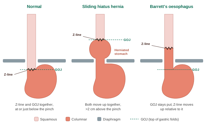
Hiatus hernia vs Barrett's: the key distinction
- Sliding hiatus hernia: the whole junction slides up. The Z-line moves up because the stomach moves up with it, so the Z-line and GOJ still coincide, but both sit more than 2 cm above the pinch.
- Barrett's oesophagus: the GOJ stays put and columnar mucosa extends up the oesophagus, so the Z-line moves up relative to the GOJ.
- Measure Barrett's (Prague C&M) from the top of the gastric folds, never from the pinch. Otherwise the hernia pouch gets mislabelled as Barrett's.
- The two often coexist, because the hernia drives the reflux that causes Barrett's.
Relevant anatomy
- The hiatus lies at about T10, slightly left of midline, and is usually formed entirely by the right crus, which splits to encircle the oesophagus. It transmits the oesophagus, both vagal trunks and oesophageal branches of the left gastric vessels.
- The phreno-oesophageal ligament (Laimer's membrane), formed from fused endothoracic and transversalis fascia, inserts 2-3 cm above the GOJ. Stretching of this ligament is the core lesion in a sliding hernia.
- The anti-reflux barrier has four parts: the intrinsic LOS (3-4 cm high-pressure zone, including sling and clasp fibres), the crural pinch, 2-3 cm of intra-abdominal oesophagus under positive pressure, and the acute angle of His acting as a flap valve. A sliding hernia weakens all four at once.
- Operative hazard: an accessory or replaced left hepatic artery, arising from the left gastric artery, runs in the pars flaccida in roughly 10-20% of people. Look before you divide it.
Classification
| Type | Description | Frequency |
|---|---|---|
| I | Sliding: GOJ above the diaphragm, stomach in its normal axis | ~90-95% |
| II | Pure paraoesophageal: GOJ in place, fundus herniates alongside | Rare |
| III | Mixed: GOJ and fundus both above the diaphragm | Most paraoesophageal hernias |
| IV | Other organs too: colon, omentum, small bowel, spleen | Uncommon |
- Types II-IV are paraoesophageal hernias (PEH). "Giant" usually means more than 30-50% of the stomach in the chest.
- Size (axial length): small under 3 cm, large 5 cm or more.
- Hill grade on retroflexion (see above): grades III-IV accompany a widened hiatus, and grade IV always has a hernia.
- High-resolution manometry (Chicago EGJ morphology): type 1 LOS and crura superimposed; type 2 separated by 1-3 cm; type 3 separated by more than 3 cm, which is a hiatus hernia with the greatest reflux burden.
Why a sliding hernia causes reflux
- The crural pinch separates from the LOS.
- The post-prandial acid pocket moves up into the hernia sac.
- Acid trapped in the sac refluxes again on swallowing ("re-reflux").
- Oesophageal clearance is impaired and transient LOS relaxations increase.
Bigger hernias mean more acid exposure, more LA C/D oesophagitis, more strictures and more Barrett's. Cameron lesions (linear erosions on the gastric folds at the level of the diaphragm) cause occult bleeding and iron-deficiency anaemia in large hernias. Sliding hernias very rarely volvulate or strangulate; that risk belongs to PEH.
Work-up before considering surgery
- OGD: hernia length, Hill grade, LA grade, Barrett's, strictures, Cameron lesions. For LA C/D, re-scope after 8 weeks of PPI.
- Contrast swallow: type, length, reducibility and rotation in large hernias. A GOJ fixed well above the diaphragm when upright suggests a short oesophagus.
- High-resolution manometry: essential before any wrap. It excludes achalasia (a disastrous thing to wrap) and absent contractility.
- Ambulatory pH or pH-impedance off PPI (stop for 7 days) if endoscopy has not already shown conclusive GORD. Lyon 2.0: conclusive GORD = LA B/C/D, long-segment Barrett's, peptic stricture or acid exposure time over 6%; normal = AET under 4%; 4-6% is borderline. A DeMeester score above 14.72 is still quoted in vivas.
- Gastric emptying study if bloating suggests gastroparesis, which a wrap would make worse.
Management: treat the reflux, not the size
Size alone is not an indication to repair a sliding (type I) hernia. Treatment is driven by the reflux and its complications.
- Asymptomatic: no treatment, no follow-up. Weight loss advice if relevant.
- GORD with no or mild oesophagitis (LA A/B): lifestyle measures (weight loss has the best evidence, head-of-bed elevation, no meals 2-3 hours before lying down, stop smoking, cut alcohol). Full-dose PPI for 4-8 weeks, then step down to the lowest effective or on-demand dose (NICE CG184). Alginates for breakthrough. PPIs treat heartburn much better than regurgitation.
- Severe oesophagitis (LA C/D), stricture or Barrett's: full-dose PPI for 8 weeks, re-scope, escalate if not healed, then long-term full-dose maintenance. Biopsy and dilate strictures. Barrett's alone is not an indication for surgery: antireflux surgery has not been shown to regress Barrett's or cut cancer risk compared with PPI.
- Consider surgery once GORD is objectively proven and:
- symptoms respond to PPI but the patient does not want lifelong medication, or cannot tolerate it;
- volume regurgitation or aspiration persists, especially with a large hernia;
- oesophagitis will not heal or strictures recur;
- extra-oesophageal symptoms (cough, laryngeal) have good symptom association, accepting a less predictable result;
- a large hernia causes mechanical symptoms or Cameron bleeding.
Good surgical candidates: typical symptoms, an abnormal pH study and a good PPI response. Poor candidates: PPI-refractory "heartburn" with normal pH (functional heartburn), atypical symptoms alone, obesity, gastroparesis and psychological comorbidity.
PEH is different: repair if symptomatic. Watchful waiting is reasonable for asymptomatic PEH, especially in older patients (emergency risk roughly 1% per year). Volvulus is an emergency: Borchardt's triad of pain, unproductive retching and inability to pass an NG tube.
Key trials
- LOTUS (JAMA 2011): laparoscopic antireflux surgery and esomeprazole gave similar remission at 5 years in PPI responders.
- REFLUX (UK; BMJ 2008, 5-year follow-up 2013): better quality of life with surgery, but about 38% of the surgical group were back on reflux medication at 5 years.
- Spechler (NEJM 2019): surgery beat medical therapy in truly PPI-refractory heartburn, but most patients screened were excluded once work-up showed their symptoms were not reflux.
Obesity: with BMI above about 35 and GORD, Roux-en-Y gastric bypass is the preferred antireflux operation. Fundoplication fails more often, and sleeve gastrectomy worsens reflux.
Alternatives: magnetic sphincter augmentation (LINX), originally for hernias under 3 cm and now used with crural repair for larger ones; it preserves belching but avoid it with nickel allergy or significant dysmotility. TIF and Stretta have limited evidence; TIF needs a hernia under 2 cm or a concurrent repair.
The operation: laparoscopic hiatus hernia repair and fundoplication
Reverse Trendelenburg, five ports, Nathanson liver retractor.
- Divide the pars flaccida, checking first for an accessory left hepatic artery.
- Identify the right crus. Open the phreno-oesophageal membrane and develop the plane outside the sac, without stripping the crural muscle.
- Create the retro-oesophageal window and preserve both vagi. The posterior vagus is most at risk here. Excise the GOJ fat pad if needed to define the junction.
- Mobilise in the mediastinum for at least 2.5-3 cm of tension-free intra-abdominal oesophagus. Too little length is the main cause of recurrence and wrap migration. If adequate length cannot be achieved, a Collis gastroplasty (stapled neo-oesophagus from the lesser curve) is needed in a small minority.
- Short gastrics: often divided for a floppy Nissen. RCTs show no clear long-term benefit for routine division, so practice varies.
- Crural repair: interrupted non-absorbable sutures posteriorly, with or without pledgets, plus anterior sutures if the axis would kink. Snug, not tight.
- Fundoplication.
| Wrap | Description | For | Against |
|---|---|---|---|
| Nissen | 360° posterior, 1.5-2 cm, floppy, over a 50-56 Fr bougie | Most durable reflux control | More early dysphagia and gas-bloat; cannot belch or vomit |
| Toupet | 270° posterior | Similar reflux control, less dysphagia and bloat | Possibly less durable |
| Dor | 180° anterior, often after Heller myotomy | Low dysphagia | Weaker reflux control |
| Anterior 90° (Watson) | Partial anterior | Low dysphagia | More long-term recurrence |
Tailoring the wrap to motility is common but not proven necessary in RCTs. Absent contractility is still a reason to avoid a 360° wrap, and to question surgery altogether.
Mesh is not routine. Oelschlager's biological mesh RCT reduced recurrence at 6 months (9% vs 24%) but not at 5 years (54% vs 59%), and Watson's Australian trial found no benefit for absorbable or non-absorbable mesh. Permanent mesh can erode into the oesophagus or stomach. Most of this data is from PEH; for sliding hernias a sutured repair is standard.
Post-op: prophylactic antiemetics (retching threatens the repair), selective contrast swallow, sloppy diet progressing over 2-6 weeks, stop PPI, no heavy lifting for several weeks.
Complications
- Intraoperative: capnothorax (usually managed conservatively), perforation, bleeding (short gastrics, spleen, liver), vagal injury, aberrant left hepatic artery injury.
- Early: dysphagia (usually settles by 6-12 weeks), acute herniation from retching, leak.
- Late: persistent dysphagia (about 5-10%), gas-bloat, inability to belch or vomit, slipped wrap (hourglass stomach), wrap migration, recurrence.
- Radiological recurrence after large hernia repair is 25-50% at 5 years, mostly small and asymptomatic. About 15-40% are back on reflux medication at 5-10 years, not all with objective reflux. Elective mortality is well under 0.5%. After repeated failure, especially in obese patients, Roux-en-Y reconstruction is often favoured.
Key numbers
| Number | Why it matters |
|---|---|
| None | No size threshold mandates repair of a type I hernia |
| 1 cm | Gastric mucosa can normally be seen up to 1 cm above the pinch |
| 2 cm | Endoscopic definition of a hernia; upper limit for TIF |
| 2.5-3 cm | Target tension-free intra-abdominal oesophagus at surgery |
| 3 cm | Original LINX limit; HRM EGJ type 3 |
| 5 cm+ | Large: predicts PPI failure and regurgitation, needs more mobilisation, recurs more |
| 4% / 6% | Acid exposure time: normal under 4%, conclusive over 6% (Lyon 2.0) |
A hernia found at fundoplication is always repaired with crural closure. For frail patients with a symptomatic large hernia, consider limited reduction with gastropexy or a PEG rather than a full repair and wrap.
12. Oesophageal and Gastric Varices
Oesophageal varices — simplified (Baveno)
Modern practice favours the simple binary:
- Small (<5 mm) — flatten with insufflation
- Large (≥5 mm) — do not flatten
The older three-grade system is still used in some units:
- Grade 1 — flattened by insufflation
- Grade 2 — not flattened, occupying <1/3 of the lumen
- Grade 3 — occupying >1/3 of the lumen
High-risk stigmata: red wale marks, cherry-red spots, haematocystic spots. These predict bleeding independently of size.
Sarin Classification — gastric varices
This one genuinely changes what you do.
| Type | Description | Management |
|---|---|---|
| GOV1 | Oesophageal varices extending along the lesser curve | Band ligation acceptable (treat as oesophageal) |
| GOV2 | Extending along the greater curve into the fundus | Cyanoacrylate glue injection |
| IGV1 | Isolated fundal varices, no oesophageal varices | Cyanoacrylate glue — and exclude splenic vein thrombosis |
| IGV2 | Isolated ectopic varices (duodenum, jejunum, colon, rectum) | Individualised |
- IGV1 with no liver disease → think splenic vein thrombosis (often secondary to pancreatitis or pancreatic malignancy). This is left-sided / sinistral portal hypertension, and it is cured by splenectomy — one of the few surgically curable causes of variceal bleeding. A classic exam question.
- Fundal varices bleed less often than oesophageal varices but bleed more severely when they do.
- TIPSS for uncontrolled or recurrent variceal bleeding.
- Terlipressin and prophylactic antibiotics for all suspected variceal bleeds — antibiotics reduce mortality.
13. Siewert Classification — Gastro-oesophageal Junction Adenocarcinoma
Defined by the position of the tumour centre relative to the anatomical GOJ.
| Type | Location | Typical surgical approach |
|---|---|---|
| I | 1–5 cm above the GOJ (distal oesophagus) | Oesophagectomy (Ivor-Lewis) |
| II | 1 cm above to 2 cm below (true cardia) | Extended gastrectomy or oesophagogastrectomy |
| III | 2–5 cm below the GOJ (subcardial gastric) | Total gastrectomy with distal oesophageal resection |
Accurate endoscopic measurement — distance from incisors to tumour, to GOJ, and to the top of gastric folds — directly determines the operation. Document it properly.
14. Zargar Classification — Caustic Oesophageal Injury
| Grade | Appearance |
|---|---|
| 0 | Normal |
| 1 | Oedema and erythema of mucosa |
| 2a | Superficial ulceration, friability, blisters, exudate |
| 2b | 2a plus deep discrete or circumferential ulceration |
| 3a | Focal necrosis (scattered black or greyish areas) |
| 3b | Extensive necrosis |
| 4 | Perforation |
Grade 2b and above predicts stricture formation. Grade 3b and 4 carry high mortality and may require oesophagectomy.
Endoscopy is best performed within 24–48 hours. Do not attempt endoscopy after 48 hours to 2 weeks — the oesophagus is at its most friable and perforation risk is highest.
15. Eosinophilic Oesophagitis — EREFS Score
Increasingly common and often missed. Remember the acronym for the endoscopic features:
- Edema (loss of vascular markings)
- Rings (trachealisation / feline oesophagus)
- Exudates (white plaques — often mistaken for candida)
- Furrows (longitudinal linear grooves)
- Strictures
Crucially: the oesophagus looks normal in up to 10–20% of cases. Any patient with dysphagia or food bolus obstruction needs biopsies from proximal and distal oesophagus (at least 6 total) regardless of appearance. Diagnosis requires ≥15 eosinophils per high-power field.
Food bolus obstruction in a young adult is eosinophilic oesophagitis until proven otherwise.
PART TWO — COLONOSCOPY
16. Boston Bowel Preparation Scale (BBPS)
Scored after washing and suctioning, for three segments (right colon, transverse, left colon).
| Score | Description |
|---|---|
| 0 | Unprepared — mucosa not seen |
| 1 | Portion of mucosa seen; staining/residue obscuring |
| 2 | Minor residual staining; mucosa well seen |
| 3 | Entire mucosa well seen |
Total 0–9.
What it changes
- Adequate preparation = total ≥6 AND every segment ≥2. A total of 6 achieved as 3+3+0 is not adequate.
- Inadequate preparation → the examination cannot be relied upon. Repeat within 1 year (many units repeat much sooner), and do not issue a normal surveillance interval on the back of a poorly prepared scope.
- Document it. Post-polypectomy surveillance recommendations assume an adequately prepared, complete colonoscopy.
17. NICE Classification (NBI International Colorectal Endoscopic)
Designed for non-magnifying narrow-band imaging — usable on standard equipment, which is why it's the practical one.
| Type 1 | Type 2 | Type 3 | |
|---|---|---|---|
| Colour | Same or lighter than background | Browner than background | Brown to dark brown, sometimes patchy white areas |
| Vessels | None, or isolated lacy vessels | Thick brown vessels surrounding white structures | Area of disrupted or absent vessels |
| Surface | Dark or white spots of uniform size, or homogeneous absence of pattern | Oval, tubular or branched white structures surrounded by brown vessels | Amorphous or absent surface pattern |
| Likely histology | Hyperplastic / sessile serrated | Adenoma | Deep submucosal invasion |
What it changes
- Type 1 — diminutive rectosigmoid lesions may be left in place or resected and discarded, depending on local policy ("resect and discard" / "diagnose and leave").
- Type 2 — resect endoscopically.
- Type 3 — do not attempt endoscopic resection. Deep submucosal invasion (>1000 µm). Tattoo adjacent to the lesion (not into it), biopsy, photograph, and refer for surgical resection and MDT discussion. Attempting piecemeal resection of a Type 3 lesion compromises subsequent surgery and pathological staging.
18. JNET Classification (Japan NBI Expert Team)
For magnifying NBI. More granular than NICE.
| Type | Vessel pattern | Surface pattern | Likely histology |
|---|---|---|---|
| 1 | Invisible | Regular dark/white spots | Hyperplastic / SSL |
| 2A | Regular calibre and distribution | Regular (tubular, branched, papillary) | Low-grade adenoma |
| 2B | Variable calibre, irregular distribution | Irregular or obscure | High-grade dysplasia / superficial submucosal invasion |
| 3 | Loose vessel areas, interruption of thick vessels | Amorphous | Deep submucosal invasion |
2B is the decision point — these need careful assessment and are best resected en bloc (ESD) or referred. Type 3 goes to surgery.
19. Kudo Pit Pattern (chromoendoscopy)
The original, using dye spray (indigo carmine, crystal violet) with magnification.
| Type | Pattern | Histology |
|---|---|---|
| I | Round pits | Normal mucosa |
| II | Stellar or papillary | Hyperplastic |
| IIIS | Small tubular/round, smaller than normal | Depressed adenoma |
| IIIL | Large tubular/round, larger than normal | Tubular adenoma |
| IV | Branched or gyrus-like | Tubulovillous adenoma |
| VI | Irregular — irregular arrangement, size, structure | High-grade dysplasia / superficial submucosal invasion |
| VN | Non-structural — loss of pit pattern | Deep submucosal invasion |
Type VN → surgery. This remains the reference standard against which newer optical classifications are validated.
20. WASP Classification — Serrated Lesions
Sessile serrated lesions are the lesions most often missed and the ones responsible for a disproportionate share of post-colonoscopy colorectal cancer. WASP combines NICE with four additional features:
- Clouded surface
- Indistinct border
- Irregular shape
- Dark spots inside the crypts
Two or more features → sessile serrated lesion.
These are typically right-sided, flat, pale, and covered by a mucus cap — wash the mucus cap off and look at the edge. They are easy to under-call and easy to resect incompletely.
21. Laterally Spreading Lesions (LSLs / LNPCPs)
LNPCP = large non-pedunculated colorectal polyp, ≥20 mm.
| Subtype | Description | Submucosal invasion risk |
|---|---|---|
| Granular, homogeneous | Uniform nodules | Low |
| Granular, mixed nodular | Nodules of varying size, dominant nodule | Intermediate — risk concentrated in the dominant nodule |
| Non-granular, elevated | Smooth surface, no nodules | Higher |
| Non-granular, pseudo-depressed | Smooth with depressed area | Highest |
Non-granular morphology and any depressed component are the red flags. Sample or resect the dominant nodule en bloc where possible.
LNPCPs should be referred to an endoscopist with the relevant expertise rather than attempted opportunistically.
22. Malignant Polyps — Haggitt and Kikuchi
Once histology shows invasive carcinoma in a resected polyp, these determine whether the patient needs a colectomy.
Haggitt levels — pedunculated polyps
| Level | Depth of invasion |
|---|---|
| 0 | Carcinoma in situ / intramucosal (not truly invasive) |
| 1 | Invades submucosa of the head |
| 2 | Invades to the neck |
| 3 | Invades the stalk |
| 4 | Invades submucosa of the bowel wall below the stalk |
Levels 1–3 have low nodal risk (~1%) if other features are favourable. Level 4 carries a significantly higher nodal risk and usually prompts consideration of segmental resection.
Kikuchi levels — sessile polyps (submucosal thirds)
| Level | Depth | Nodal metastasis risk |
|---|---|---|
| sm1 | Upper third (<1000 µm) | ~1–3% |
| sm2 | Middle third | ~8% |
| sm3 | Lower third | ~23% |
The features that actually drive the decision
Regardless of Haggitt or Kikuchi level, consider surgical resection if any unfavourable feature is present:
- Poor differentiation (grade 3)
- Lymphovascular invasion
- Tumour budding (grade 2–3)
- Resection margin <1 mm (or involved)
- Submucosal depth ≥1000 µm
Absence of all of these = favourable = endoscopic resection alone is likely sufficient, with surveillance.
This is an MDT decision balancing nodal risk against operative mortality — an sm1 lesion with favourable features in an 85-year-old is managed very differently from the same lesion in a 55-year-old.
23. Post-Polypectomy Surveillance — BSG/ACPGBI/PHE 2020
The 2020 guidelines abolished the old high/intermediate/low-risk three-tier system. There are now only two outcomes: surveillance or discharge. Consultants who trained on the 2002/2010 guidelines sometimes still quote the old intervals — know the difference.
High-risk criteria → one-off colonoscopy at 3 years
Any one of:
- ≥2 premalignant polyps including ≥1 "advanced" polyp, where advanced means:
- Adenoma ≥10 mm or containing high-grade dysplasia, or
- Serrated polyp ≥10 mm or containing any grade of dysplasia
- ≥5 premalignant polyps
Everyone else → discharge to the national bowel cancer screening programme
Note the counterintuitive consequence: a single isolated 15 mm adenoma does not meet the criteria for surveillance. This surprises people, and it is a favourite question.
Other pathways
| Situation | Follow-up |
|---|---|
| Post-CRC resection | Clearance colonoscopy at 1 year, then surveillance colonoscopy 3 years later |
| Piecemeal EMR of LNPCP (≥20 mm) | Site check at 2–6 months, further site check at 18 months, then 3-yearly surveillance per high-risk criteria |
| Incomplete index colonoscopy or poor prep | Repeat before assigning any interval |
Subsequent surveillance is determined by applying the same high-risk criteria to the findings at each surveillance procedure.
24. Inflammatory Bowel Disease
24a. Mayo Endoscopic Subscore — Ulcerative Colitis
| Score | Appearance |
|---|---|
| 0 | Normal or inactive disease |
| 1 | Mild — erythema, decreased vascular pattern, mild friability |
| 2 | Moderate — marked erythema, absent vascular pattern, friability, erosions |
| 3 | Severe — spontaneous bleeding, ulceration |
- Mucosal healing was historically defined as 0 or 1; the trend, and the target in most modern trials, is Mayo 0.
- Simple, universally used, but has poor inter-observer agreement at the 1/2 boundary.
24b. UCEIS — Ulcerative Colitis Endoscopic Index of Severity
More reproducible. Three descriptors scored at the most severely affected area:
- Vascular pattern (0–2): normal / patchy obliteration / obliterated
- Bleeding (0–3): none / mucosal / luminal mild / luminal moderate-severe
- Erosions and ulcers (0–3): none / erosions / superficial ulcer / deep ulcer
Total 0–8. Remission ≤1.
24c. Montreal Classification — extent
Ulcerative colitis:
- E1 — proctitis (limited to rectum)
- E2 — left-sided (distal to splenic flexure)
- E3 — extensive / pancolitis (proximal to splenic flexure)
Crohn's disease — Age at diagnosis (A1 <17, A2 17–40, A3 >40); Location (L1 ileal, L2 colonic, L3 ileocolonic, L4 upper GI); Behaviour (B1 non-stricturing non-penetrating, B2 stricturing, B3 penetrating, p perianal modifier).
24d. SES-CD — Simple Endoscopic Score for Crohn's Disease
Four variables scored 0–3 across five segments (ileum, right colon, transverse, left colon, rectum):
- Size of ulcers
- Ulcerated surface
- Affected surface
- Presence and type of narrowing
Total 0–56. Remission <3. Mucosal healing = complete absence of ulceration.
24e. Rutgeerts Score — Post-operative Crohn's Recurrence
Assessed at the neoterminal ileum at colonoscopy 6–12 months after ileocolonic resection. This one is surgically essential.
| Score | Findings |
|---|---|
| i0 | No lesions |
| i1 | ≤5 aphthous ulcers |
| i2 | >5 aphthous ulcers with normal intervening mucosa, or skip areas, or lesions confined to the ileocolonic anastomosis (<1 cm) |
| i3 | Diffuse aphthous ileitis with diffusely inflamed mucosa |
| i4 | Diffuse inflammation with large ulcers, nodules, and/or narrowing |
i0–i1 = remission. i2 and above = significant endoscopic recurrence → escalate medical therapy.
The critical point: endoscopic recurrence precedes clinical recurrence by years. Nearly all patients have endoscopic recurrence within a year of resection, and the Rutgeerts score at 6–12 months predicts symptomatic and surgical recurrence. This is why post-operative colonoscopy is mandatory and why patients should not be discharged after their operation.
(Note: i2 is sometimes subdivided into i2a — lesions confined to the anastomosis, and i2b — >5 aphthous lesions in the neoterminal ileum. Some units treat only i2b and above as true recurrence.)
24f. IBD Dysplasia Surveillance
- Chromoendoscopy with targeted biopsies is preferred over random biopsies.
- Interval determined by risk: 1, 3 or 5 years depending on extent, disease activity, family history of CRC, presence of PSC, and previous dysplasia.
- Primary sclerosing cholangitis → annual colonoscopy from diagnosis, regardless of other factors.
- Dysplasia should be classified as visible (endoscopically resectable or not) or invisible — this determines whether resection or colectomy is appropriate.
25. Goligher Classification — Haemorrhoids
Clinic rather than endoscopy, but you will be asked in the same breath.
| Grade | Description | Typical management |
|---|---|---|
| I | Bleed but do not prolapse | Conservative: fibre, fluids, topical |
| II | Prolapse on straining, reduce spontaneously | Rubber band ligation |
| III | Prolapse, require manual reduction | Band ligation or surgery (HALO, haemorrhoidectomy) |
| IV | Irreducible / permanently prolapsed | Haemorrhoidectomy |
Always exclude a proximal cause of bleeding before attributing rectal bleeding to haemorrhoids — particularly in anyone over 40, with altered bowel habit, iron deficiency anaemia, or a family history. Haemorrhoids are common; so is coexisting colorectal cancer.
26. Colonoscopy Quality Indicators (BSG/JAG)
Worth knowing — they come up in interviews, appraisal, and audit.
| Indicator | Minimum standard | Aspirational |
|---|---|---|
| Caecal intubation rate (unadjusted) | 90% | 95% |
| Adenoma detection rate | 15% | 20% |
| Polyp detection rate | 15%+ | — |
| Polyp retrieval rate | 90% | — |
| Rectal retroflexion | 90% | — |
| Terminal ileal intubation (suspected IBD) | 60% | — |
| Patient comfort | <10% moderate–severe discomfort | — |
| Withdrawal time (normal colonoscopy) | ≥6 minutes | — |
- Photodocumentation of the caecum (ileocaecal valve and appendiceal orifice, or terminal ileum) is required as proof of completion.
- JAG certification requires a minimum of 280 colonoscopies (200 if already certified in flexible sigmoidoscopy), 15+ procedures in the preceding 3 months, and completion of the JAG Basic Skills course.
- Every additional 1% increase in ADR is associated with approximately a 3% reduction in interval cancer risk — the strongest argument for slow, careful withdrawal.
QUICK REFERENCE — What Your Consultant Is Actually Asking
| They point at… | They want… | And then… |
|---|---|---|
| Oesophageal erosions | LA grade | C/D → repeat OGD after 8 weeks PPI |
| Salmon-pink mucosa | Prague C&M | Seattle protocol biopsies, interval by length |
| Bleeding ulcer | Forrest class | Ia/Ib/IIa → dual-modality therapy + PPI infusion |
| Z-line above the pinch | Hiatus hernia (Z-line and GOJ together, >2 cm above) vs Barrett's (Z-line above the GOJ) | Hernia: treat the reflux, not the size. Barrett's: Prague C&M from the top of the folds |
| A non-bleeding gastric ulcer | Benign vs malignant features, Johnson type | Rim biopsies, H. pylori, PPI 8 weeks, repeat OGD at 6-8 weeks |
| A duodenal ulcer | Cause | H. pylori biopsies, PPI 4-8 weeks, no repeat OGD |
| Dyspepsia, normal-looking stomach | H. pylori status | Antral biopsies (plus body if on PPI), state H. pylori on the form |
| Iron-deficiency anaemia | A complete look | Sydney gastric biopsies + D2 biopsies for coeliac |
| Red stripes in the antrum | GAVE vs portal hypertensive gastropathy | GAVE → APC |
| A flat/depressed lesion | Paris type | Any IIc or III component → be very cautious |
| Red/atrophic gastric mucosa | Kyoto / Kimura and Sydney biopsies | OLGIM III–IV → 3-yearly surveillance |
| Fundal varices | Sarin type | IGV1 → glue + exclude splenic vein thrombosis |
| GOJ tumour | Siewert type | Determines the operation |
| A colonic polyp | Paris + NICE type | NICE 3 → tattoo, don't resect, refer |
| Right-sided pale flat lesion | WASP | ≥2 features → sessile serrated lesion |
| Bowel prep | BBPS | <6 or any segment <2 → repeat |
| UC mucosa | Mayo / UCEIS | Guides escalation |
| Neoterminal ileum post-op | Rutgeerts | ≥i2 → escalate therapy |
| Polyps just removed | BSG 2020 criteria | ≥2 with 1 advanced, or ≥5 → 3 years. Otherwise discharge |
A Note on Using This
Classifications are shorthand for a management decision. When you are asked one on the list, give the grade and the consequence — "LA grade C, so I'd want to repeat after eight weeks of PPI to exclude underlying Barrett's" lands considerably better than "grade C."
Where UK and European guidance differ (Barrett's surveillance intervals, most obviously), say so. Knowing that a difference exists is a stronger answer than confidently quoting one of them.
This reference reflects guidance current as of October 2026. BSG, ESGE and NICE guidance is periodically revised — verify against current published guidance and local trust protocol before applying to patient care. This document is educational and is not a substitute for clinical judgement.
For educational use only. Always check your local trust protocol and the current guideline position before acting on anything here.